SYNOVIAL SARCOMA
- Hits: 1575
Synovial sarcoma is a rare high-grade malignant soft tissue tumor. Although synovial sarcoma can be seen in many different age groups, unlike other soft tissue sarcomas, it often occurs in young adults (15-35 years).
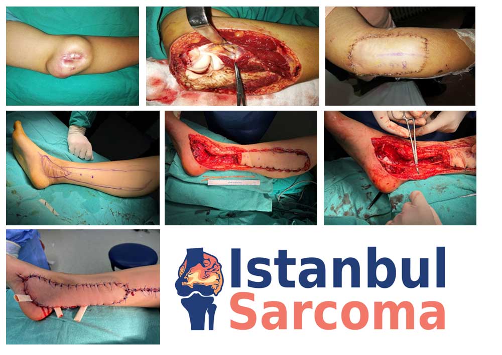
Synovial sarcomas are often found around joints (synovial bursa, tendon sheath and fascia, etc.) such as the knee, elbow and foot.
Despite their name, they do not originate from intra-articular synovial tissue and have different types (monophasic, biphasic, epithelioid).
Patients diagnosed with synovial sarcoma often present with swelling. Compared to other soft tissue sarcomas, synovial sarcomas are smaller in size and may be associated with pain.
Unlike other soft tissue sarcomas, synovial sarcomas are slow growing and small in size. The patient may have a long medical history. This may mislead the patient and physician into believing that it is benign.
Physical examination reveals a soft and mobile mass. Unlike other soft tissue sarcomas, the mass may be painful to palpation.
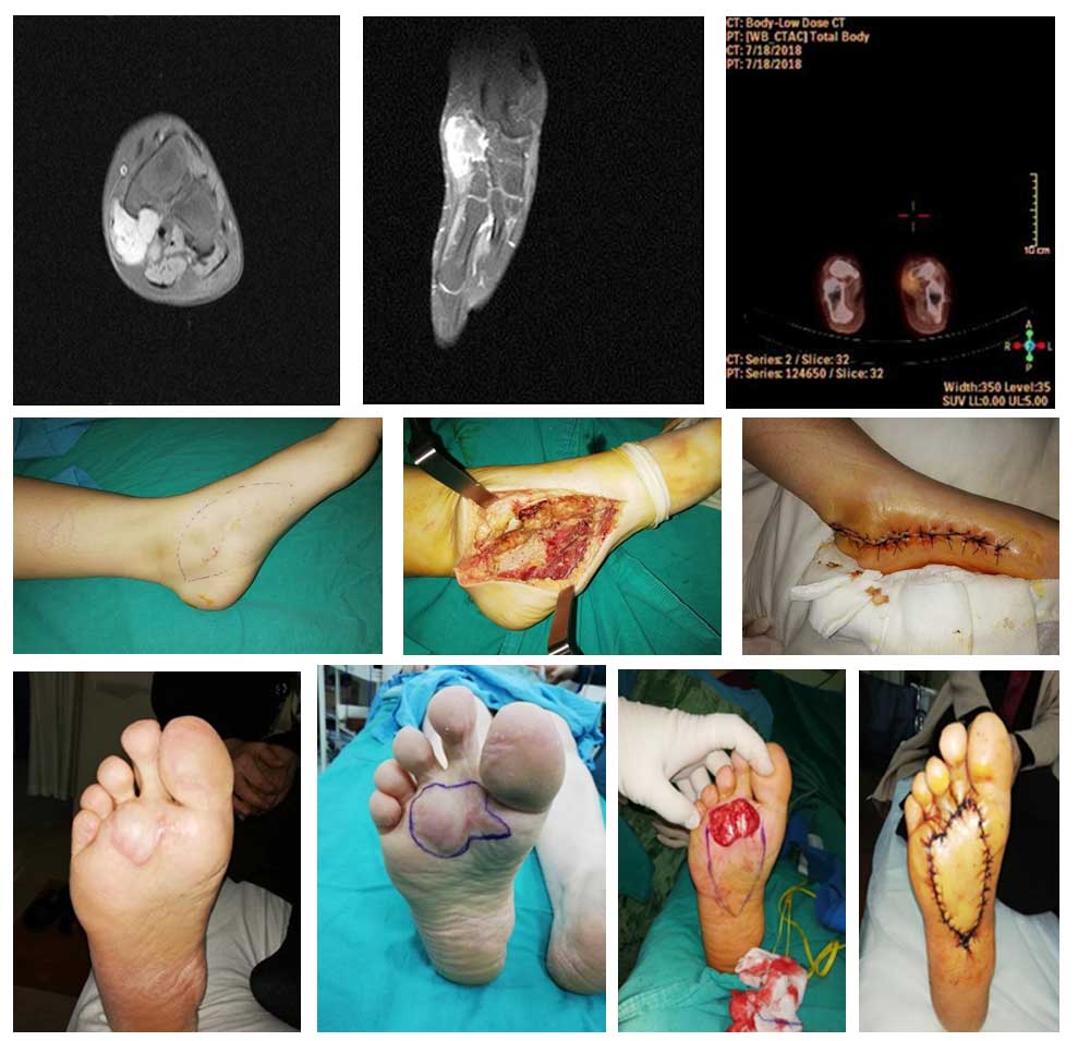
The first imaging modality to be performed in synovial sarcoma is x-ray, and calcifications can be seen on x-ray. This image is important for diagnosis. In these patients, we most often use MRI with medication as the imaging method. MRI gives us detailed information about the location, size, boundaries, and contents of the tumor. MRI is also used to plan surgery, evaluate response to chemotherapy or radiation, and monitor for recurrence.
The definitive diagnosis of synovial sarcoma is made by biopsy after clinical and radiologic evaluation. The biopsy is usually performed in a closed procedure under local anesthesia using special needles. It is important that the physician who performs the biopsy is an orthopedic oncologist who specializes in bone and soft tissue tumors, and that the pathologist who evaluates the biopsy sample is experienced in this field.
Patients diagnosed with synovial sarcoma will undergo a CT scan, whole-body MRI or PET-CT to detect metastases (lymph nodes, lungs). Synovial sarcoma is prone to metastasis via the lymph nodes.
The primary treatment for synovial sarcoma is surgical removal of the tumor in a wide, clean fashion. The tumor that is not removed cleanly with wide margins has a nearly one hundred percent recurrence rate and is prone to metastasis. For this reason, it is important that the surgeon who performs the surgery is an orthopedic oncologist with experience in this field.
Radiotherapy is used before or after surgery to facilitate synovial sarcoma surgery and reduce the possibility of recurrence. Although there are advantages and disadvantages to using radiation before or after surgery, there is no absolute superiority of one over the other.