SPINE-VERTEBRAE TUMORS
- Hits: 473
Tumors of the spine (vertebrae) can be defined in several ways. First, it is necessary to distinguish between tumors of the vertebral bone and tumors of the spinal cord. Here we will talk about tumors of the vertebral bone.
Vertebral tumors are divided into two groups: tumors that originate from their own tissue (primary) and tumors that originate from other sites of cancer (metastatic/secondary).
The spine is the third most common site where cancer cells metastasize, after the lungs and liver. Most tumors that affect the spine have spread (metastasized) to the spine from elsewhere in the body, usually from the prostate, breast, lung, kidney, thyroid, or colon. More than 40% of patients with cancer anywhere in the body will have their cancer spread to the spine during their lifetime. Fortunately, only 10% of patients will experience symptoms. For this reason, vertebral tumors are more common in people with a history of cancer.
Tumors that originate from the spine's own tissue (primary) are much less common. Primary tumors may be benign or malignant.
- A benign spinal tumor usually has smooth edges. A benign tumor is not cancerous, but it may need to be removed if it is causing symptoms, damaging the spine, or pressing on nerves. Examples include aneurysmal bone cyst, osteoid osteoma, osteoblastoma, osteochondroma, and hemangioma.
- Malignant tumors are cancers that can spread to adjacent healthy tissues, invade other organs (lungs, liver, etc.), and be life-threatening. Malignant vertebral tumors include chondrosarcomas, Ewing sarcomas, and osteosarcomas.
Vertebral tumors can cause a variety of symptoms, especially as they grow and damage the tissues where they are located. In addition to your spinal bone, tumors may affect your spinal cord or nerve roots, blood vessels, or nearby organs. Signs and symptoms of vertebral tumors may include the following
- The most common complaint is back pain. Back pain is not relieved by rest and can be worse at night.
- Loss of sensation or muscle weakness, especially in the arms or legs
- Difficulty walking, sometimes leading to falls
- Loss of bowel or bladder function (inability to urinate or defecate / loss of control)
- Paralysis may develop slowly or rapidly, affecting part or all of the arm or leg.
- Vertebral tumors progress at different rates depending on the type of tumor.
When to see a doctor
There are many causes of back pain, and most back pain is not caused by a tumor. However, because early diagnosis and treatment are important for vertebral tumors, tell your doctor the following information about your back pain:
- Persistent and progressive
- Not related to a specific activity
- Worse at night
- A history of cancer and recent back pain
Seek medical attention immediately in the following cases:
- Progressive muscle weakness or numbness in your legs or arms
- Changes in bowel or bladder function
Prognosis
The prognosis of a vertebral tumor depends on whether the tumor is benign or malignant. Benign tumors usually heal well with appropriate surgery. For malignant tumors that originate in the spine itself (primary), a good prognosis depends on the tumor not spreading to other organs, complete surgical removal, and a good response to chemotherapy and/or radiation therapy.
For vertebral tumors that originate from another cancer site (metastases), the prognosis depends mainly on the type of cancer present, the presence of spread to organs other than the spine, and the general condition of the patient.
Risks
Both benign and malignant vertebral tumors can compress the nerves in your spinal cord, causing a loss of movement or sensation below the site of the tumor. This can sometimes cause changes in bowel and bladder function. Nerve damage can be permanent.
A vertebral tumor can also damage the bones of the spine and make them unstable, increasing the risk of a sudden fracture or collapse of the spine that could injure the spinal cord.
However, if the tumor is detected early and treated quickly, it may be possible to prevent further loss of function and restore nerve function. Depending on its location, a tumor that presses against the spinal cord itself can be life-threatening.
Diagnosis
Vertebral tumors can sometimes be overlooked because their symptoms are similar to those of more common conditions. Therefore, it is important that your doctor obtain a complete medical history and perform a general physical and neurological examination.
If your doctor suspects a vertebral tumor, one or more of the following tests may help confirm the diagnosis and locate the tumor:
Plain x-ray: Plain x-rays are used to show erosion of the pedicles or vertebral body. However, radiographic findings are not apparent until 30-50% of the bone has been destroyed.
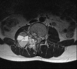
- Magnetic Resonance Imaging (MRI). MRI uses a powerful magnet and radio waves to create precise images of your spine, spinal cord, and nerves. MRI is often the test of choice for diagnosing vertebral tumors. A contrast dye, which helps make certain tissues and structures more visible, may be injected into a vein in your foot or forearm during the test.
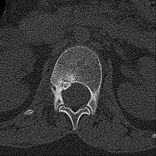
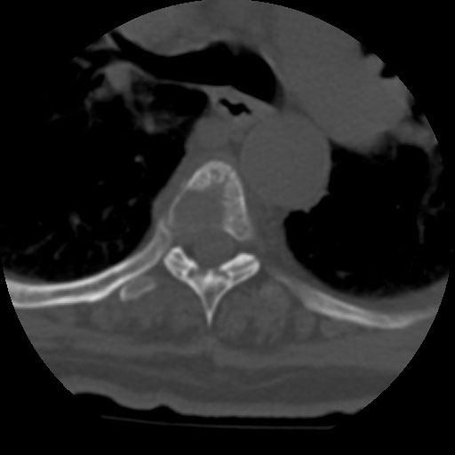
- Computed Tomography (CT). While MRI shows soft tissues in greater detail, CT is superior and more useful in showing bony structures. CT may be used in combination with MRI. In addition, in patients with metastases of unknown origin, CT scans of the lungs and abdomen are performed to identify the primary focus.
In metastatic patients, imaging should include the entire spine due to the possibility of lesions in other spinal locations (15%).
Bone scan (scintigraphy): Used to confirm whether there is a lesion in a bone other than the spine, especially in patients with metastases.
Positron emission tomography (PET)-CT: Allows for rapid screening and staging of systemic disease. It may also be used as a control in cancer patients to determine response to chemotherapy.
- Biopsy. Often, the only way to determine the type of tumor is to take a small sample of tissue (biopsy) and examine it under a microscope. The results of the biopsy will help determine treatment options.
The method used to obtain the biopsy sample can be critical to the success of the overall treatment plan. In most cases, the procedure is performed under anesthesia with an imaging (usually X-ray or CT) guided closed procedure with a biopsy needle.
Treatment options for most vertebral tumors include:
- Surgery.
Surgical removal of the vertebral tumor and filling of the cavity: Ideally, the goal of vertebral tumor treatment is to remove the tumor completely. However, there is a risk of permanent damage to the spinal cord or surrounding nerves. This option is usually preferred for patients who are in good general health and have a long life expectancy.
Removal of tumor tissue causing spinal cord compression and stabilization the spine: Tumors, especially malignant tumors, can cause compression of the spinal cord, resulting in partial or complete paralysis. In this case, the spinal cord is drained to relieve the compression (decompression surgery) and the affected spine is stabilized with screws (instrumentation).
Vertebroplasty/Kyphoplasty: These procedures reconstruct collapsed vertebral bone to correct alignment or relieve pressure on a nerve. Vertebroplasty and kyphoplasty are image-guided procedures performed in an operating room with x-ray equipment (scopes).
This procedure is performed under either general anesthesia or local anesthesia-assisted sedation (where the patient is awake). Under x-ray guidance, one or two needles are inserted through the back skin into the fractured vertebrae. After confirming good needle placement with scopy, the surgeon injects bone cement into the fractured vertebra. The cement hardens within minutes, providing immediate bone stability and pain relief. This method can be used in cancer patients with poor general condition.
- Radiotherapy: This may be used after surgery to remove remnants of tumors that cannot be completely removed or to treat inoperable tumors.
It may be the first-line treatment for some spinal tumors. Radiation therapy may also be used to relieve pain only when surgery is too risky.
Stereotactic radiosurgery (SRS). This treatment, which is not actually surgery, delivers high doses of precisely targeted radiation. With SRS, doctors use computers to focus radiation beams with pinpoint accuracy at all points and from multiple angles. It is highly effective and has fewer side effects than conventional radiotherapy. In radiosurgery, there are several types of technologies (Cyberknife, etc.) that use radiation stereotactically to treat vertebral tumors.
- Chemotherapy: A standard treatment for many types of cancer, chemotherapy is used to destroy cancer cells or stop them from growing. It may be used alone or in combination with other therapies.
- Hormonotherapy: It is commonly used in patients with prostate and breast cancer.
- Immunotherapy: Used in patients with metastatic melanoma, lung cancer and kidney cancer
- Steroids: Surgery and radiation therapy, as well as the tumors themselves, can cause non-microbial inflammation in the spinal cord, and doctors sometimes prescribe corticosteroids to reduce edema after surgery or during radiation therapy.
Although corticosteroids reduce inflammation, they are usually used only for a short time because they cause serious side effects such as muscle weakness, osteoporosis, high blood pressure, diabetes, and increased susceptibility to infection.
- Bisphosphonates: These bone-strengthening drugs reduce the risk of collapse and fracture of tumor-affected bones.