RHABDOMYOSARCOMA
- Hits: 735
Rhabdomyosarcoma is the most common malignant soft tissue tumor in children. It is less common in adults.
It has a rapid and aggressive progression. It can metastasize to the lungs, brain, and bones via the hematogenous route. It also tends to metastasize by lymphatic route. The 5-year survival rate is approximately 65%.
They often occur in the head-neck and abdomen-pelvis regions, rarely in the extremities.

Rhabdomyosarcoma originates from striated muscle and has different subtypes (embryonal, alveolar, pleomorphic and mixed).
- Embryonal: This is the most common type. It is common in males under the age of 10. 10% occur in the extremities. The tumor is soft in consistency.
- Alveolar type: It is seen in the extremities and paravertabral area between 10-25 years of age. It is a hard tumor and may show multiple localizations. The prognosis is poor.
- Pleomorphic type: This is the rarest type and has the worst prognosis. It is a fast growing, painful, hard mass with indistinct borders that tends to localize in the lower extremities (thigh-hip) in middle-aged and elderly patients. It is rarely seen in the upper extremity (arm).
Patients with rhabdomyosarcoma often present with swelling. Pain is rarely present.
The tumor is usually deep and large. It may be located in the arms and legs or in the abdomen and pelvis.
Physical examination of patients with rhabdomyosarcoma often reveals a painless, mobile, soft mass.
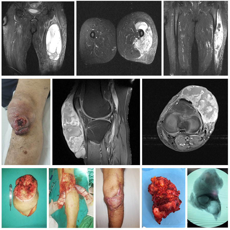
For patients with rhabdomyosarcoma, we most commonly use MRI with medication as the imaging modality. MRI gives us detailed information about the location, size, boundaries and contents of the tumor. MRI is also used in planning surgery, evaluating response to chemotherapy and radiation, and monitoring for recurrence.
The definitive diagnosis of rhabdomyosarcoma is confirmed by biopsy after clinical and radiologic evaluation. The biopsy is often performed using a closed procedure with special needles. It is important that the physician who performs the biopsy is an orthopedic oncologist who specializes in bone and soft tissue tumors, and that the pathologist who evaluates the biopsy sample is experienced in this area.
Patients diagnosed with rhabdomyosarcoma will undergo a CT scan, whole-body MRI, or PET-CT to look for metastases (usually in the lungs).
Rhabdomyosarcoma is more sensitive to chemotherapy and radiation than other malignancies. Therefore, treatment consists of chemotherapy, radiation therapy, and surgical removal. Chemotherapy and radiotherapy may be given before or after surgery. For surgical treatment, clean removal of the tumor with wide margins is recommended. Tumors that are not cleanly removed with wide margins have a nearly one hundred percent recurrence rate and are prone to metastasis. For this reason, it is important that the surgeon performing the surgery be an orthopedic oncologist with experience in this area.
Patients diagnosed with rhabdomyosarcoma should be followed for many years at regular intervals for recurrence and metastasis.

