")
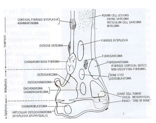
Destruction Type (Lodwick)
- Geographical pattern (type I)
- Piecemeal pattern (type II)
- Permeative pattern (type III)
English, Русский, O‘zbekcha, Türkçe
![]()
![]()
Malignant bone tumors are very rare compared to benign ones. Usually, patients seek with pain, and sometimes with swelling. We are faced with a delay in the diagnosis of such tumors because the tumor should take time to grow enough to cause complaints, it is not important because it causes mild complaints at first, and it does not show any findings in roentgen graphy taken at an early stage. The fact that there is pain that wakes you up from sleep at night is an important sign for suspecting these tumors. Chondrosarcoma is usually diagnosed in adult patients, while osteosarcoma and Ewing's sarcoma occur in childhood and young adulthood.
According to the location:
The diagnosis of such patients is made by a biopsy, which is performed in addition to clinical and radiological evaluation. Although blood tests do not make a definitive diagnosis, they can support our diagnosis (Alkaline phosphatase and Lactic dehydrogenase height) and are used in the differential diagnosis from other lesions such as bone infection (CRP height). Radiologically, direct graphy (simple x-ray) is a must-have and medicated (contrast-enhanced) MRI is one of our routine examinations. We perform the biopsy procedure under anesthesia in the operating room with the help of a needle, often closed, accompanied by imaging (scopy). An experienced bone pathologist plays a vital role in evaluating the biopsy taken. Additional bone scintigraphy and lung tomography techniques are required for staging (whether there are other bone or lung metastases) from patients who have been diagnosed.
A patient who has been diagnosed and screened is referred to medical oncology and given chemotherapy. (Chemotherapy is effective only for some types of chondrosarcoma patients while routine chemotherapy is given in osteosarcoma and Ewing's sarcoma) Control X-ray and MRI are taken from the patient who has finished chemotherapy and the surgery is performed. During the surgery, the cancerous bone is removed and replaced with a prosthesis, as well as the cavity can be filled with bone taken from himself/herself (fibula), or the removed bone with tumor is cleaned and put in place with an implant/plaque after undergoing some procedures (liquid nitrogen or radiotherapy). After the surgery, the patient whose wound site has healed is referred to the medical oncology unit for chemotherapy again.
Teşvikiye Mah. Hakkı Yeten Cad.
Doğu İş Merkezi , No:15 Kat:7
Şişli, İstanbul
English, Russian, Uzbek, Türkçe
Medical Coordinator
Is there anything I can help you with?.
09.00