English, Русский, O‘zbekcha, Türkçe
![]()
![]()
BENIGN BONE TUMORS AND CYSTS
Bone tumors are rare lesions. A large part of these tumors are benign lesions, and they often occur by chance during radiological examinations for another reason. As a complaint, they usually show swelling and pain. Rarely, it is diagnosed after causing a fracture in the place where it was. Osteoid osteoma, osteochondroma (exostosis), enchondroma, non-ossifying fibroma, eosinophilic granuloma, simple bone cyst, fibrous dysplasia and intraosseous lipoma/ganglion are the most common lesions of benign tumors.
Clinical Detections of Benign Bone Tumors and Cysts
- Most of the cases occur in childhood and adolescence.
- It is consulted with complaints of swelling and pain.
- Rarely, the diagnosis is made after a fracture formation.
- Some of them are detected by chance.
- It usually has limited growth capacity.
- Treatment is not always required, some just follow up.
Classification of Benign Bone Tumors and Cysts (Enneking)
Classification of benign bone tumors and cysts according to the tissue from which they originate
- Stage 1
They are self-limiting lesions that are usually diagnosed by chance; Osteochondroma, Enchondroma, Non-Ossifying Fibroma (NOF), Fibrous dysplasia, Eosinophilic granuloma and Simple bone cyst - Stage 2
Lesions with limited destruction in the surrounding tissue, often manifested by pain; Osteoblastoma, Chondroblastoma, Chondromyxoid fibroma. - Stage 3
They are lesions with an aggressive character, the boundaries of which are not very clear; Aneurysmal bone cyst, Giant cell tumor.
Classification of benign bone tumors and cysts according to the tissue from which they originate
VISUALISATION OF BONE TUMORS
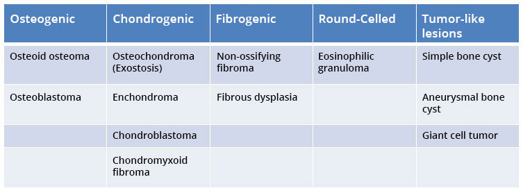
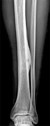
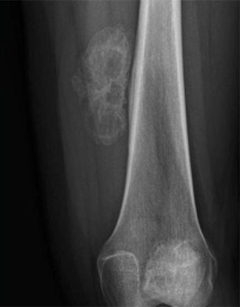
DIRECT GRAPHY
The first step is a direct graphy (X-ray). A pre-diagnosis of many patients can be made by direct graphy.
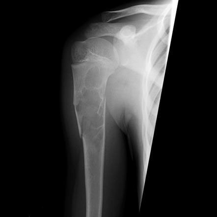
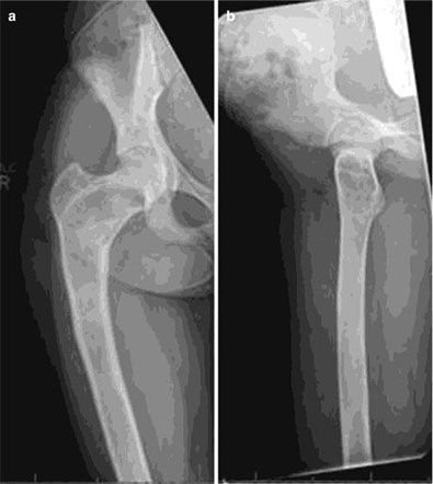
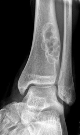
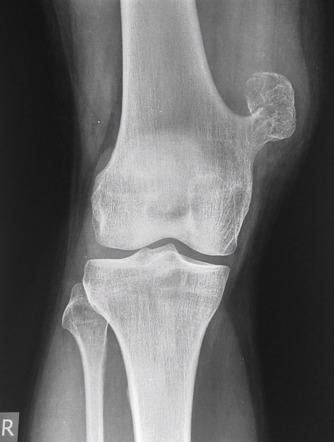
EXAMPLES OF DIRECT GRAPHY
COMPUTED TOMOGRAPHY (CT)
-
- Shows the bone details better.
- In some tumors, it is a direct diagnostic tool. (Osteoidosteoma)
EXAMPLES OF COMPUTED TOMOGRAPHY (CT)
 ÖRNEKLERİ")
MAGNETIC RESONANCE (MRI)
-
- It is superior in showing the soft tissue component accompanying bone tumors.
- We evaluate the relationship of the tumor with vascular structures.
- It helps to decide where the biopsy will be performed.
- It is routinely taken medicated (contrast-enhanced).
EXAMPLES OF MAGNETIC RESONANCE (MRI)
")
Benign bone tumors
- Simple bone cyst
- Aneurysmal bone cyst
- Osteoid osteoma
- Osteoblastoma
- Enchondroma
- Osteochondroma (Exostosis)
- Chondroblastoma
- Fibrous Dysplasia
- Non-ossifying fibroma
- Eosinophilic granuloma
- Giant cell tumor
Malignant tumors that can be confused with benign tumors
-
- Aneurysmal bone cyst - Telangiectatic osteosarcoma
- Osteoblastoma - Low-grade osteosarcoma
- Eosinophilic granuloma - Lymphoma, Ewing's sarcoma
- Osteochondroma, periosteal chondroma-parosteal osteosarcoma
- Giant cell tumor - Osteosarcoma rich in giant cells

Address
Teşvikiye Mah. Hakkı Yeten Cad.
Doğu İş Merkezi , No:15 Kat:7
Şişli, İstanbul