CHONDROSARCOMA
- Hits: 799
Chondrosarcoma is a malignant tumor of the bone that originates from cartilage tissue. Chondrosarcoma usually occurs after the age of 40 and is more common in men.
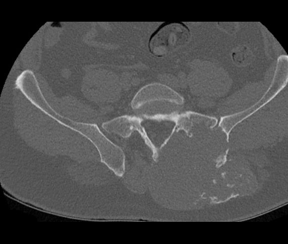
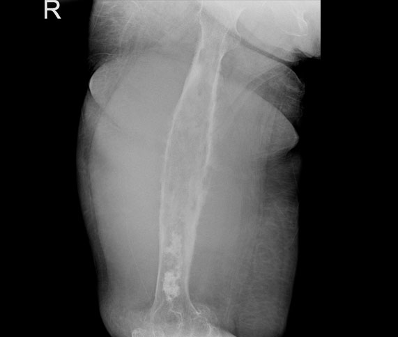
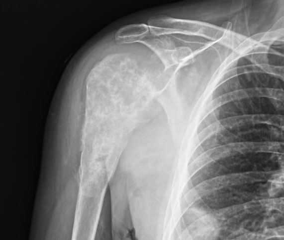
Chondrosarcoma is most commonly found around the pelvis, hip (proximal femur), and shoulder (scapula, proximal humerus). It is often found in the metaphyseal region of long bones.
Chondrosarcomas most commonly occur in bone, but rarely in soft tissue (extraskeletal chondrosarcoma).
Chondrosarcomas develop in two forms: primary (most common) and secondary (on the background of pre-existing benign cartilage tumors such as enchondromas and osteochondromas). Patients with multiple enchondromatosis (Olier, Mafucci syndrome) and hereditary osteochondromatosis are at increased risk. Therefore, these patients should be closely monitored for malignant transformation. Although there are many subtypes of primary chondrosarcomas, the most common types are classical (grades I, II, III), followed by mesenchymal, myxoid, and dedifferentiated types.
The most common finding in patients with chondrosarcoma is pain and swelling. The pain may be mild at first, but may increase over time to become severe, persistent at rest, and not responsive to pain medication. Swelling is often hard and immobile and may be tender. In addition, neurological and vascular symptoms may develop due to compression of the mass.
In patients with chondrosarcoma, we should start imaging methods with a direct radiograph (X-ray). X-rays give us a lot of information about the location, size, internal structure and character of the tumor. We especially use tomography to get information about bone destruction and Magnetic Resonance Imaging (MRI) to evaluate the soft tissue part and the boundaries of the tumor. We also use MRI for surgical treatment planning. A large and heterogeneous cartilage tumor with cortical destruction and soft tissue extension should be a warning sign for chondrosarcoma.
There is no significant value in laboratory tests in chondrosarcoma patients.
In patients with suspected chondrosarcoma, we resort to biopsy to confirm the diagnosis of the patient whose clinical and radiologic evaluation is complete. Although biopsy can be performed with closed needle or open methods, the open method comes to the fore especially in cartilage-derived tumors. It is important that the physician who performs the biopsy is an orthopedic oncologist who specializes in bone and soft tissue tumor surgery, and that the microscopic examination is performed by a physician who specializes in bone and soft tissue pathology.
Patients diagnosed with chondrosarcoma should be evaluated for metastatic disease. Tomography, whole-body MRI, scintigraphy, and PET-CT may be used for screening.
The main treatment for chondrosarcoma is surgery. In surgery, the tumor is removed with clean margins (intralesional intervention can be performed in grade 1 chondrosarcoma) and the resulting gap is reconstructed with biological (graft, allograft, fibula, etc.) or non-biological (prosthesis) methods. Tumors that are not removed cleanly with wide margins have a recurrence rate of almost one hundred percent and are prone to metastasis. For this reason, it is essential that the surgeon performing the surgery is an experienced orthopedic oncologist.
Chemotherapy is ineffective in classical chondrosarcoma but may be used in dedifferentiated and mesenchymal types. Radiation therapy is ineffective and is not used in routine treatment.
Patients should be followed for many years after chondrosarcoma surgery, especially for recurrence and metastasis.