GIANT CELL TENDON TUMOR - PIGMENTED VILLONODULAR SYNOVITIS
- Hits: 432
It is a benign but locally aggressive tumor. In other words, it can cause bone and joint destruction in the area where it is located and has a high risk of recurrence after surgery.
The exact cause is unknown. They can be intra-articular or extra-articular. There are two types: localized and diffuse.
The diffuse type used to be called pigmented villonodular synovitis.
The diffuse intra-articular form usually involves single and large joints, and the knee joint is most commonly affected. After the knee, the hip and ankle are most commonly affected.
30-50 years of age
Recurrent pain, swelling, and limitation of movement are the most common reasons for referral to a physician. Bloody serosanguineous fluid may be found on aspiration for joint swelling.
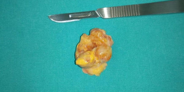
Because of its slow progression, many years may pass before a diagnosis is made. During this time, arthritis may develop due to cartilage destruction. While the tumor may be very large in the diffuse type, it usually does not exceed 3-4 cm in the localized type.
Joint destruction, subchondral cysts and calcifications may be seen on direct radiography. MRI is the gold standard for diagnosis. It is also helpful in planning surgical treatment and follow-up. Biopsy is not routinely used in diagnosis. In doubtful cases, it may be performed to confirm the diagnosis. Biopsy is usually performed as a closed needle biopsy under local anesthesia. It is important that the physician who performs the biopsy is an orthopedic oncologist who specializes in bone and soft tissue tumors, and that the pathologist who evaluates the biopsy sample is experienced in this area.
Synovial sarcoma, rheumatoid arthritis, and synovial hemangioma are important in the differential diagnosis.
For intra-articular localizations, open or arthroscopic excision can be performed depending on the size and location of the tumor. For tumors located in the knee, surgery can be performed through two separate incisions, anterior and posterior. The effectiveness of the treatment depends on the quality of the synovectomy. Cauterization of the tumor bed after synovectomy is recommended. Intralesional curettage may be used for bone or joint destruction. In very extensive and surgically risky masses, radionuclide synovectomy may be performed by injecting Yttrium-90 into the joint. Depending on the degree of joint destruction, prosthetic reconstruction may be a good option.
There is a high recurrence rate of up to 40% after surgery. For this reason, it is recommended that an orthopedic oncologist specializing in bone and soft tissue tumors perform the treatment, especially for diffuse types. In recurrent cases, surgical excision is performed again.
Chemotherapy and radiotherapy are not used in routine treatment, but radiotherapy and targeted therapies may be used especially in diffuse recurrence and very large tumors.